Artificial Scab Formation with a CO₂ Laser: A Summary of a 2023 Review

CO₂ lasers are widely recognized in dentistry for soft-tissue cutting and hemostasis. A 2023 review published in Diseases, however, examined another potential role: using laser-assisted blood coagulation to help create an environment that supports the healing of small defects in oral soft tissue.
The paper, titled CO₂ Laser for Esthetic Healing of Injuries and Surgical Wounds with Small Parenchymal Defects in Oral Soft Tissues, identifies two factors that may contribute to esthetic and functional healing: artificial scab formation and photobiomodulation therapy, or PBMT.
This article expands on our short video introduction to artificial scab formation. The video provides a concise visual overview, while the sections below take a closer look at the mechanisms and clinical applications discussed in the review.
This first part of our research summary focuses on what the authors describe as an “artificial scab,” how it is formed, and the clinical applications discussed in the review.
Wound Healing and Scar Formation
Small injuries and surgical wounds in oral soft tissue do not always heal with the original tissue contour fully preserved. Depending on the size, depth, and location of the tissue defect, healing may involve scar formation, contracture, or a concavity in the epithelial surface. These changes may affect both appearance and function, particularly in mobile areas such as the lips, cheeks, and tongue.
The review briefly distinguishes between primary and secondary wound healing.
Primary healing generally occurs when wound edges are brought together and closed with sutures. Properly aligned suturing can promote early wound closure. However, the authors note that tension, contraction, or dead space beneath the wound may contribute to scar formation or changes in the surrounding tissue.
Secondary healing occurs when a wound remains open. Blood coagulates at the wound surface, granulation tissue develops within the defect, and epithelium gradually extends over the area. During this process, myofibroblasts help contract and close the wound, but that contraction can also contribute to scarring and changes in tissue contour.
Wound dressings are often used to support secondary healing. They help retain blood and wound fluid, maintain moisture, and protect developing epithelial and granulation tissue. In oral tissues, however, movement and moisture can make it difficult to secure and maintain a conventional dressing.

The Artificial Scab Described in the Review
The authors use the term “artificial scab” to describe a layer of coagulated and carbonized blood formed on the wound surface through high-intensity laser therapy, or HILT.
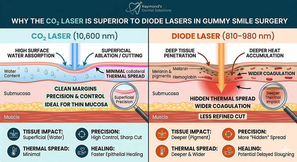
The paper explains that the 10.6 μm wavelength of a dental CO₂ laser is strongly absorbed by water. When blood is irradiated under appropriate conditions, coagulation and carbonization occur primarily at the surface, while the blood beneath that surface layer remains.
Rather than removing or absorbing all the blood from the wound, the technique described in the review allows blood to accumulate within the tissue defect. The surface is then coagulated to form the artificial scab, which is attached, or “soldered,” to the surrounding mucosa to reduce the likelihood of separation.
The authors describe three principal roles for this layer.
Maintaining Space Within the Wound
The review refers to a “space-making effect.” By covering blood and wound fluid retained within the defect, the artificial scab may help preserve space for tissue repair and regeneration.
This concept is particularly important when the objective is not only to close a wound, but also to maintain the original shape or volume of the affected tissue.
Preserving a Moist Healing Environment
Blood and wound fluid contain components involved in cell migration, division, and proliferation. According to the review, retaining these fluids beneath the artificial scab helps create a moist wound-healing-like environment.
In this respect, the artificial scab is described as having a function similar to a wound dressing.
Protecting the Wound Surface
The authors also describe the artificial scab as a covering that protects immature epithelial and granulation tissue from contamination and infection.
They suggest that it may avoid some practical limitations of conventional dressings because it is breathable, separates naturally rather than requiring removal, and can be attached to surrounding tissue in highly mobile areas.
Clinical Applications Discussed in the Paper
The review presents four principal applications involving artificial scab formation. These examples illustrate how the same underlying concept may be applied differently depending on the anatomy and type of wound.

Mucocele Removal
In the technique described for mucocele removal, a CO₂ laser is used to cut and vaporize tissue. After the lesion is removed, blood is retained within the resulting space and shaped along the contour of the lip. An artificial scab is then formed over the blood and attached to the surrounding tissue.
The paper explains that this approach generally does not require suturing. However, the authors also note that detailed evidence regarding long-term esthetic outcomes, motor function, and scar formation remains limited.
Open Lip Wounds
For open wounds of the lip vermilion, the review emphasizes preserving the lip contour while minimizing unnecessary removal of tissue. Following debridement, blood is allowed to accumulate within the defect, and an artificial scab is formed at the surface.
This application requires particular care. The authors warn that forming an artificial scab without properly aligning the wound edges may result in irregular closure, concavity, unevenness, or hypertrophic scar formation. Light suturing may therefore be needed to align the edges before the artificial scab is created.
Extraction Sockets
The paper also discusses artificial scab formation after tooth extraction. Blood is allowed to fill the socket to the level of the surrounding mucosa, and the blood surface is coagulated with a CO₂ laser.
The authors propose that this covering helps prevent surrounding epithelium from extending into the extraction socket and helps maintain space for bone regeneration. The biological explanation presented in this section draws partly from the authors’ previous animal studies, including histological observations in rat extraction sockets.
The review therefore presents an interesting space-maintenance concept, but it should not be interpreted as definitive clinical proof that the technique produces superior human extraction-site outcomes.
Free Gingival Graft Donor Sites
At a free gingival graft donor site, an artificial scab may serve a function similar to that of a periodontal pack or artificial dermis. Blood is retained at the exposed donor site, and its surface is coagulated to provide wound coverage.
The authors state that research involving CO₂ lasers in this application remains very limited. They also explain that they use a scalpel, rather than a CO₂ laser, to harvest the gingival graft because thermal protein coagulation at the graft margins could affect the tissue.
Interpreting the Evidence Carefully
This publication is a review article, not a new randomized clinical trial. It brings together basic research, animal studies, clinical studies, and clinical examples to describe how artificial scab formation may support the healing of selected oral soft-tissue wounds.
The authors acknowledge that research involving scars in oral mucosa is limited compared with research involving skin. In addition, artificial scab formation is followed by PBMT in many of the procedures described in the paper. This makes it difficult to separate the contribution of the artificial scab from the effects of PBMT.
Appropriate case selection and laser technique are also essential. The review specifically warns that excessive irradiation caused by unsuitable output, irradiation mode, exposure time, distance, or operator movement can result in irreversible tissue changes and delayed healing.
A Broader View of Laser-Assisted Hemostasis
The review presents artificial scab formation as more than a method of stopping bleeding. In the procedures described by the authors, coagulated blood becomes a wound covering intended to retain blood and wound fluid, maintain space, preserve moisture, and protect the healing surface.
This concept offers a different perspective on the role of CO₂ laser-assisted coagulation in oral soft-tissue procedures. At the same time, the available evidence remains limited, and further comparative clinical research is needed to determine how these techniques perform relative to conventional wound-management methods.
In Part Two, we will continue reviewing the paper’s second major focus: how CO₂ laser PBMT may influence wound healing and scar formation.
Source
Daigo Y, Daigo E, Fukuoka H, et al. CO₂ Laser for Esthetic Healing of Injuries and Surgical Wounds with Small Parenchymal Defects in Oral Soft Tissues. Diseases. 2023;11(4):172. https://doi.org/10.3390/diseases11040172




Comments